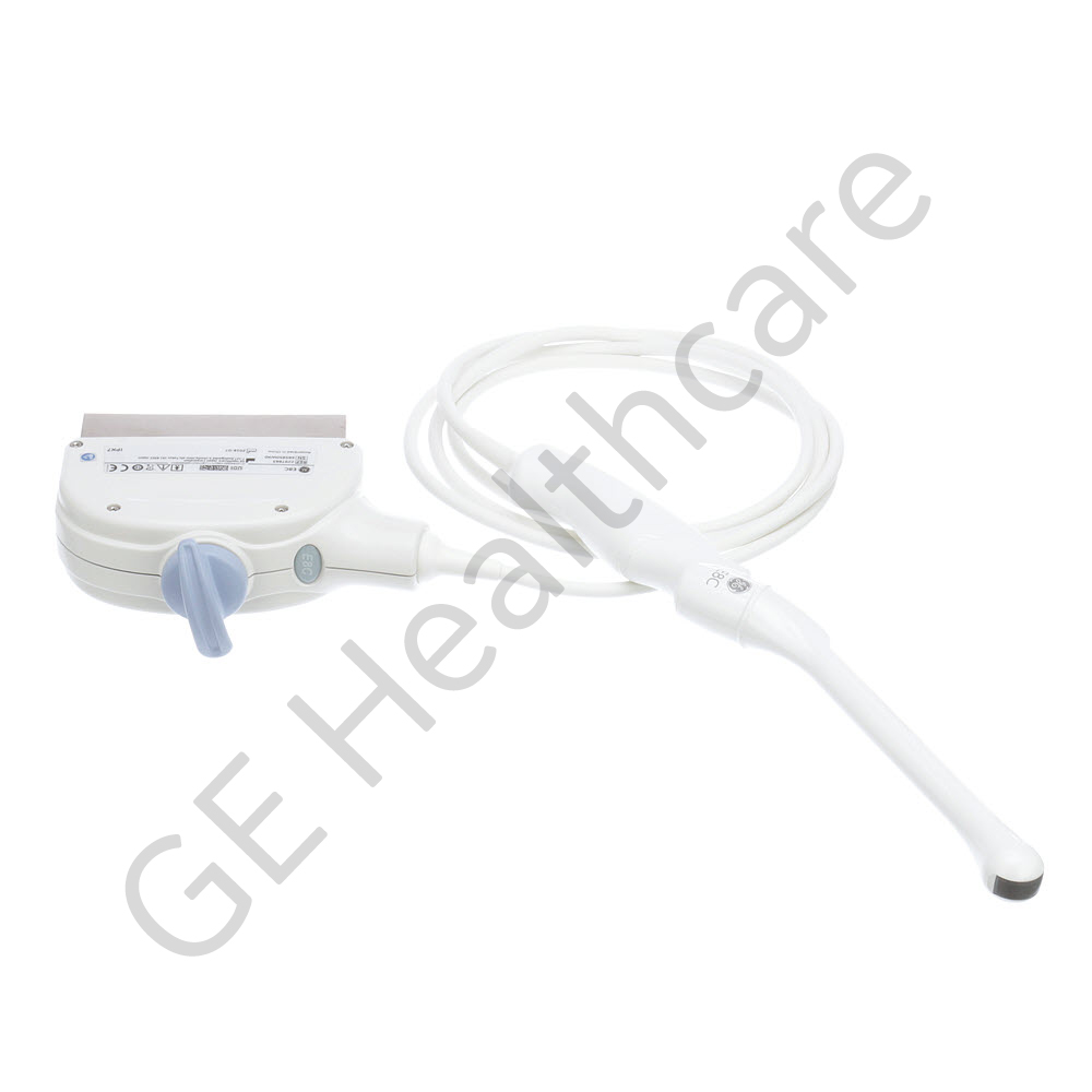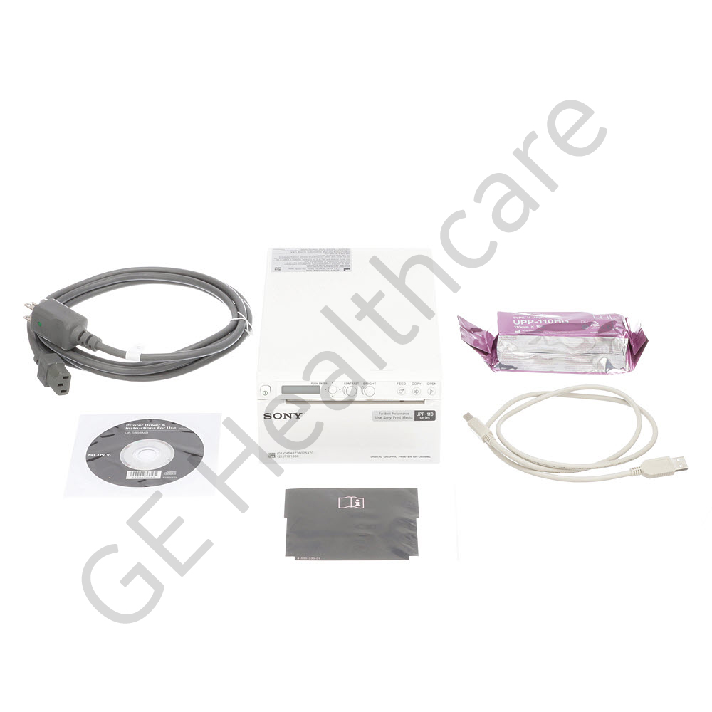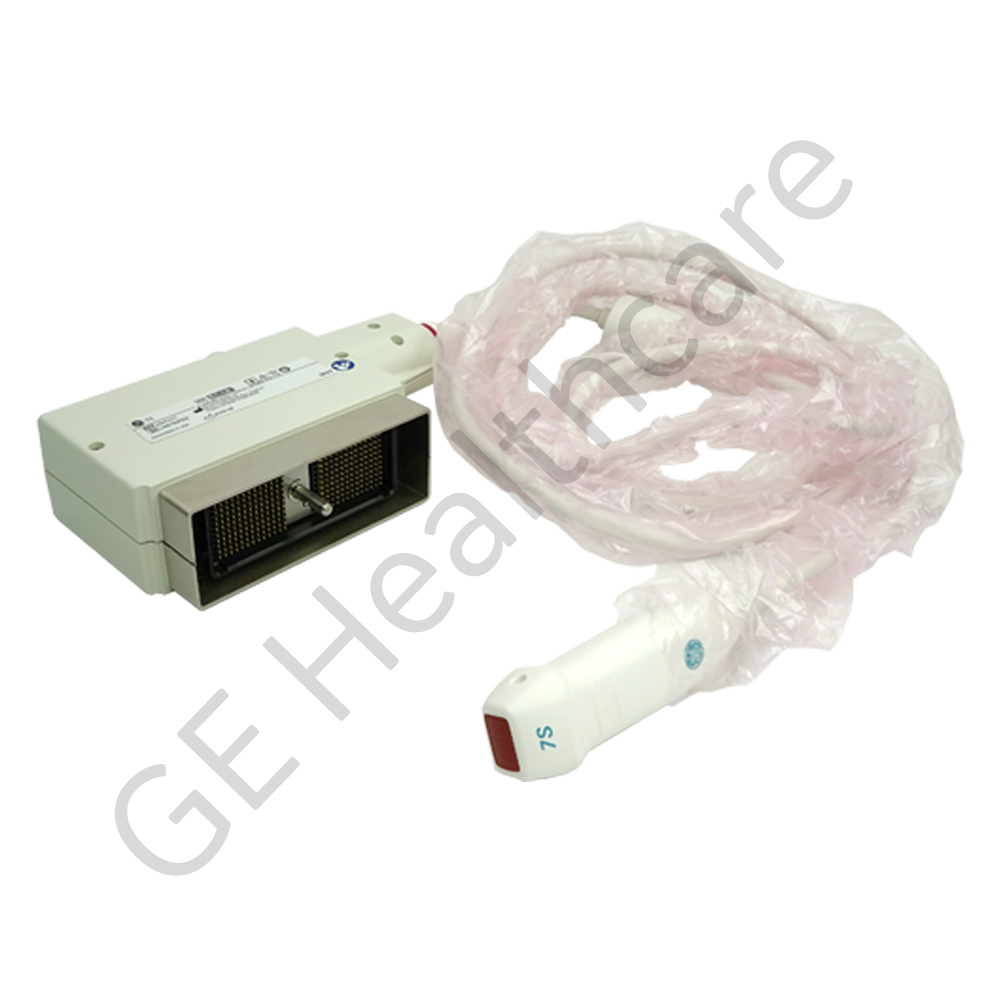

Collector, Non-RoHS, Non-CE Mark Probe, 7S
| 5505552 | |
| 2263669 2347471 | |
| New | |
| GE HealthCare | |
| GE HealthCare | |
| GE HealthCare | |
| N/A | |
Enter your approval number and submit to add item(s) to cart.
Please enter approval number
OR
Don't know your approval number? Call 800-437-1171
Enter opt 1 for the first three prompts, and have your System ID available.
If you add item(s) to cart and submit your order without the
approval number, GE will contact you before your order
can be confirmed for shipment.
Select your approver's name and submit to add item(s) to your cart
Please Select Approver Name
OR
Don't know your approval number? Call 800-437-1171
Enter opt 1 for the first three prompts, and have your System ID available.
If you add item(s) to cart and submit your order without
selecting an approver, GE will contact you before your order
can be confirmed for shipment.
Features
- Comfort scan design
- Ease of use
- Ergonomic benefits
- Patient and user comfort
Frequently purchased with this item
Product Overview
Collector, Non-RoHS, Non-CE Mark
Additional Features
- High precision in dimensional measurement
- Compact design
- Can be used for Cardiac, Abdomen
- High accuracy
Compatible Products

Probes
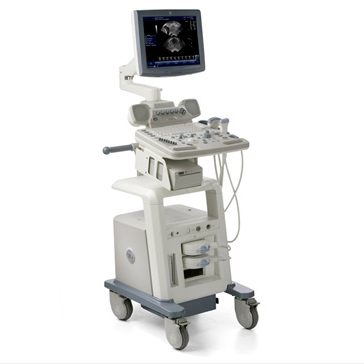
LOGIQ P5
The LOGIQ* P5 portable ultrasound system puts quality ultrasound technology well within the reach of your private practice, specialized clinic, or hospital. It offers solid image quality, including enhanced 4D capabilities and an extensive portfolio of transducers to help you take your patient care to a high level.
Advanced features help you capture exceptional images:
High Definition Speckle Reduction Imaging (SRI-HD) helps eliminate noise while maintaining true tissue architecture
CrossXBeam* Imaging helps enhance tissue and border differentiation
Auto Optimization (AO) increases contrast resolution
Auto TGC helps provide a homogenous brightness to the image to enhance clarity
Phase Inversion Harmonics helps provide higher spatial resolution and deeper penetration
3D/4D imaging helps reveal anatomical details
Elastography for breast and musculoskeletal exams
Auto IMT (Automated Intima Media Thickness) for quick carotid artery analysis
TVI, Myocardial Doppler imaging with color overlay
Stress Echo
LOGIQ P5 includes many established applications from GE's high-end ultrasound systems, including:
A broad portfolio of broadband transducers
Enhanced 4D capabilities
Intuitive image management tools

LOGIQ P6
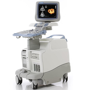
Vivid 7
The Vivid 7/Vivid 7 PRO ultrasound unit is a high performance digital ultrasound imaging system.The system provides image generation in 2D (B) Mode, Color Doppler, Power Doppler (Angio), M-Mode, Color M-Mode, PW and CW Doppler spectra, Tissue Velocity imaging and Contrast applications. The fully digital architecture of the Vivid 7/Vivid 7 PRO unit allows optimal usage of all scanning modes and probe types,throughout the full spectrum of operating frequencies.The Vivid 7 Dimension gives clinicians a better way to convey their findings to other cardiologists, referring physicians,EP physicians and patients. Now, cardiac anatomy,synchronicity and viability can be clearly communicated in imaging formats that are more familiar for your clinical partners, thus easier to understand.
• Real-time 4D imaging – provides more cardiac information to help clinicians better communicate the heart’s structure and function.
• 4D Tissue Synchronization Imaging (TSI) – propels Tissue Velocity Imaging (TVI) to the next level by taking three simultaneous planes – from a single heartbeat at high frame rates – to create a flexible, dynamic 4D model with quantitative measurements to better communicate cardiac dyssynchrony.
• Bull’s-eye report formats and TSI surface mapping –communicate cardiac dyssynchrony in a visual display that should be more familiar to EP physicians.
• Blood Flow Imaging (BFI) – new vascular imaging mode gives clinicians a better understanding and delineation of directional blood flow in vessels.
• Seamless measurement integration – allows you to efficiently calculate ejection fraction and volumes from tri-plane images gathered from the same heartbeat.
Equivalent Item(s):
Below is more information on the equivalent item(s). Items without a hyperlink are listed for reference only and are not available for purchase online.
| Equivalent Item(s) | Item Details |
|---|---|
| 2263669 | 7S PROBE_COLLECTOR |
| 2347471 |
|


